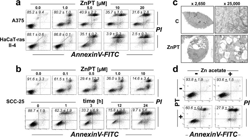
Figure 2. ZnPT-induced cytotoxicity in malignant human skin cell lines.
Dose response of ZnPT-induced cell death. Cells were exposed to ZnPT (≤ 20 µM; 24 h) or left untreated, and viability was assessed by flow cytometry [annexin V-PI staining; numbers in quadrants indicate viable (AV-negative, PI-negative) in percent of total gated cells. (a) A375; HaCaT-ras II-4. (b) SCC-25 cells: ZnPT dose response (upper panel), time course analysis (lower panel; ZnPT; 10 µM). (c) Transmission electron microscopy (fold magnification as indicated) using SCC-25 cells to ZnPT (10 µM; 6 h). (d) SCC-25 cells were exposed to the isolated or combined treatment of zinc acetate (25 µM) and pyrithione (5 µM). Cell viability was then assessed as in panel (a).

