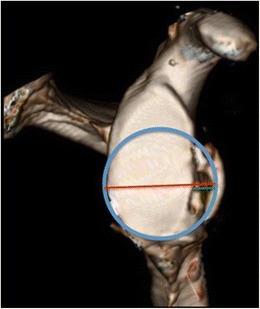
Fig. 3.

3D-MRI of glenoid, en face view, demonstrating the best-fit circle method of quantifying glenoid bone loss. (Reproduced, with permission, from Gyftopoulos S, Beltran LS, et al. use of 3D MR reconstructions in the evaluation of glenoid bone loss: a clinical study. Skeletal Radiol. 2014 Feb;43(2):213–8)
