FIGURE 3.
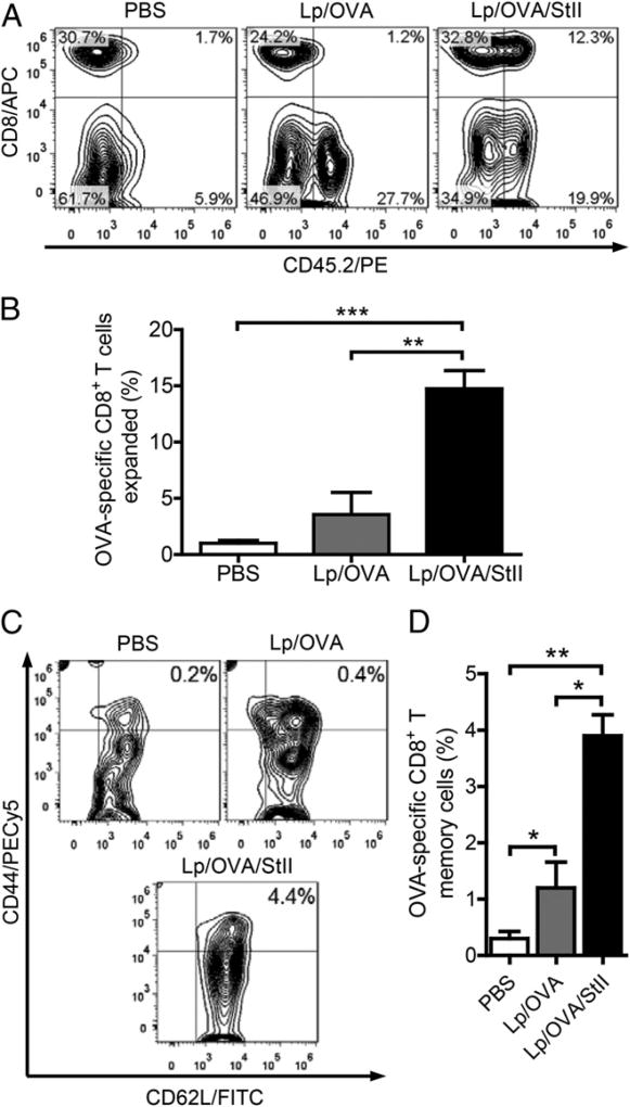
Immunization with StII coencapsulated into the DPPC:Cho Lp with OVA induces significant expansion of OVA-specific CD8+ T lymphocytes. Splenocytes (25 × 106 equivalent to 5.5 × 10⁶ OVA-specific CD8+ T cells) from OT-1 mice (CD45.2+) were transferred i.v. into Ly5 mice (CD45.1+). Two days later, Ly5 mice (n = 3) were immunized s.c. twice (12-d interval) with Lp/OVA/StII (50 µg OVA and 6.25 µg StII), Lp/OVA (50 µg OVA), or PBS (as negative control). Seven days after the second immunization, the inguinal LN closest to the inoculation site of each mouse was removed and homogenized, and CD8+ CD45.2+ cells were analyzed by flow cytometry. (A) Each contour graph, with the respective cell percentages in each quadrant referred to the lymphocyte gate, corresponds to an individual animal representative of each group. (B) Percentage (mean ± SEM) of CD8+ CD45.2+ cells for each treatment. (C) Contour graphs showing the percentage of CD44high CD62L+ cells (CD8+ memory cells) in relation to the lymphocyte gate of an individual mouse representative of each immunized group. Graphs are from the CD8+ CD45.2+ gate. (D) Percentage (mean ± SEM) of CD44high CD62L+ cells for each group. Data in (B) and (D) are from a meta-analysis of two independent assays. *p < 0.05, **p < 0.01, ***p < 0.001, two-tailed unpaired t test.

