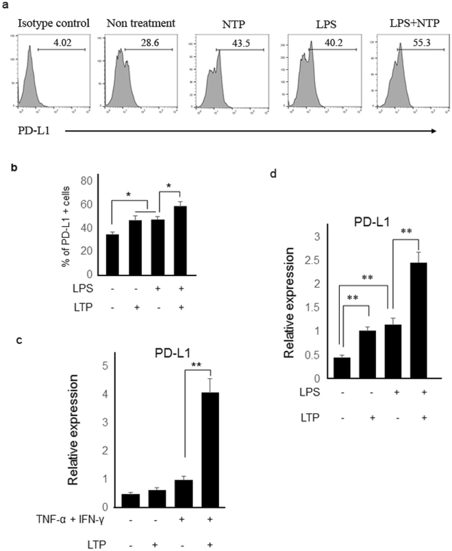
Figure 6.
Plasma treatment enhanced PD-L1 expression in HaCaT cells. (a) LPS-stimulated and LTP-stimulated HaCaT cells induced higher PD-L1 expression than did the untreated HaCaT cells. LPS/LTP-stimulated HaCaT cells had more increased expression of PD-L1 than LTP only- or LPS only-stimulated cells. (b) The percentage of PD-L1 positive cells are indicated as a graph. Transcript levels of PD-L1 were also determined by real-time PCR. LTP treatment into (c) TNF- α/IFN-γ-stimulated or (d) LPS-stimulated HaCaT cells enhanced PD-L1 gene expression. Three independent experiments were performed. *P < 0.05, **P < 0.005.

