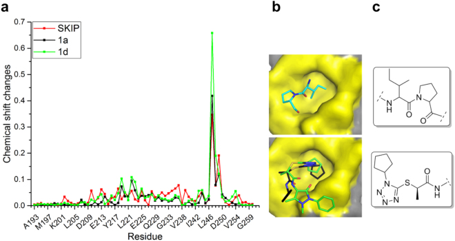
Figure 3.
Comparison between the IP mimetic compounds and the natural SxIP motif. (a) Chemical shift changes plot for SKIP peptide (red), 1a (black) and 1d (green). (b) 3D model for the IP motif and the IP motif mimetic. The IP motif three dimensional representation is based on the crystal structure 3GJO. The IP mimetic compounds 1a (black) and 1d (green) are shown in the binding poses predicted by our docking studies using 3GJO structure as the EB1c model. In both representations the C-terminus tail was removed for clarity. (c) 2D structure of IP motif and IP mimetic scaffold.

