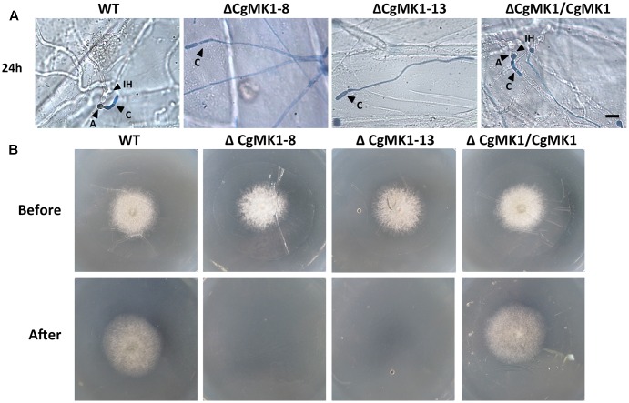
FIGURE 6.
Assays for penetration and colonization defects in the CgMK1 mutants. (A) Onion epidermal cell penetration assay of the ΔCgMK1 mutants. The assay was performed by inoculating conidial suspensions from the indicated strains. Examination under a light microscope was performed after aniline-blue staining. A: appressorium; C: conidium; IH: invasive hyphae. Scale bar: 10 μm. (B) Cellophane membranes penetration assay was performed by the conidial suspensions from strains. Colonies of the indicated strains grown on PDA plates covered with a cellophane membrane (before). Then the medium was removed the cellophane membrane and placed for additional days (after). Photos in the first row were taken at 3 dpi, the second row were taken at 5 dpi that showed growth of strains after penetration from cellophane membrane.

