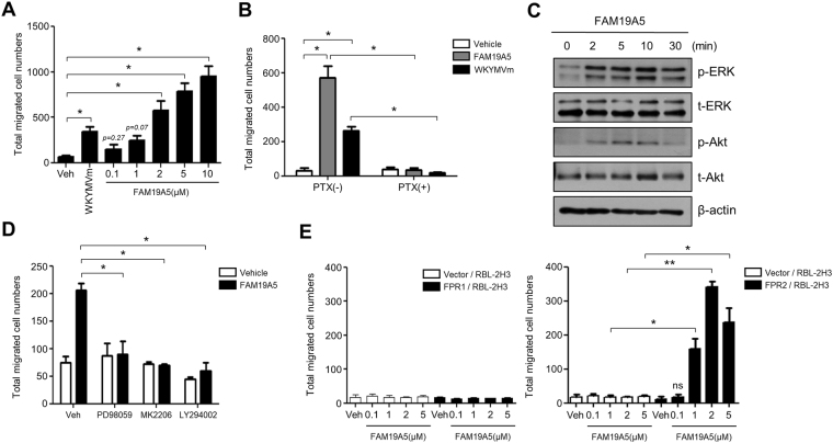
Figure 1.
FAM19A5 stimulates BMDM chemotaxis via FPR2. (A) Mouse BMDMs were used for chemotaxis assay using multiwell chamber containing several concentrations (0, 0.1, 1, 2, 5, 10 μM) of FAM19A5 or 1 μM of WKYMVm for 2 h. (B) Mouse BMDMs were incubated in the absence or presence of 500 ng/ml PTX for 4 h and applied to the upper well of the multiwell chamber containing 2 μM of FAM19A5 or 1 μM of WKYMVm for 2 h. (C) Mouse BMDMs were stimulated with 2 μM of FAM19A5 for 0, 2, 5, 10, and 30 min. Total cell lysates were separated by SDS-PAGE. Levels of p-ERK and p-Akt were measured by Western blot analysis. Data are representative of three independent experiments (C). (D) Mouse BMDMs were incubated in the absence or presence of PD98059 (50 μM) for 60 min, LY294002 (50 μM) for 15 min, or MK-2206 (2 μM) for 20 min and applied to the upper well of the multiwell chamber containing 2 μM of FAM19A5 for 2 h. (E) Vector-, FPR1-, or FPR2- expressing RBL-2H3 cells were applied to the upper well of a multiwell chamber containing 2 μM of FAM19A5 for 4 h. The number of migrated cells was determined by counting under a light microscope (A,B,D,E). Data are presented as means ± SE (n = 3). Data are representative of at least three independent experiments. ns: not significant, *p < 0.05; **p < 0.01.

