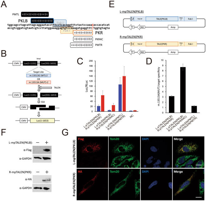
Figure 3.
Generation of G13513A-mpTALEN. (A) Schematic illustration of target site of G13513A-pTALEN. Black and white boxes indicate RVDs of L-pTALEN and R-pTALEN, respectively. Letters beside box indicate TALE’s name. Black bar indicates spacer region of L-pTALEN(PKLB)/R-pTALEN(PKR) pair. (B) Scheme of SSA assay. The reporter plasmid encodes a part of the mtDNA sequence including m.13513G or m.13513A, named m.13513 G(WT)-2 or m.13513A(MUT)-2, which is sandwiched by two split inactive parts of the luciferase gene with overlapping repeated sequences. Following a double strand break caused by TALENs, a functional luciferase gene is generated by an SSA reaction. (C) Evaluation of SSA activity (Luc/RLuc) of pTALEN pairs. Blue and red bars reflect cleaving activity against m.13513 G(WT)-2 and m.13513A(MUT)-2, respectively. NC, negative control. Data are expressed as mean SEM (n = 3). (D) m.13513A(MUT) target specificity of each L-pTALEN/R-pTALEN pair. Data are expressed as mean SEM (n = 3). (E) Components of plasmids used to express L-mpTALEN(PKLB) and R-mpTALEN(PKR) monomers. (F) Western blotting of lysed HEK293T cells transfected with plasmid coding L-mpTALEN(PKLB) or R-mpTALEN(PKR) for 2 days. L-mpTALEN(PKLB) or R-mpTALEN(PKR) was detected using anti-Flag or anti-HA antibodies, respectively. GAPDH serves as a loading control. (G) Intracellular localization of mpTALENs analyzed by immunocytochemical analysis. L-mpTALEN(PKLB) or R-mpTALEN (PKR) was transiently expressed in HeLa cells. Two days after transfection, L-mpTALEN(PKLB) or R-mpTALEN(PKR) was stained using anti-Flag or anti-HA antibodies, respectively (red). Mitochondria were stained using anti-TOM20 antibodies (green). Nuclei were stained with DAPI (blue). Scale bar, 20 μm.

