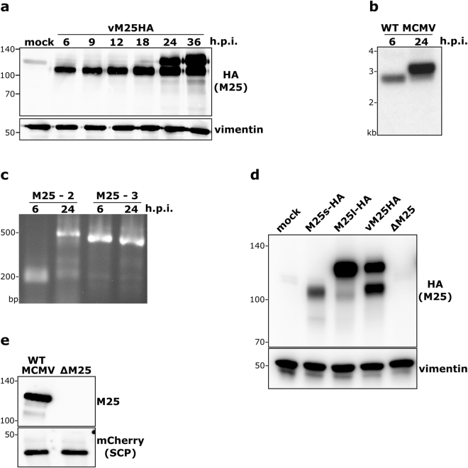
Figure 2.
Transcripts and proteins originating from the M25 ORF. (a) Lysates of MEF either mock-infected or infected with the vM25HA virus (at MOI 1) and harvested at the indicated time points were subjected to immunoblotting with an HA antibody. Vimentin served as loading control. Protein size markers (in kDa) are indicated to the left. (b) Total RNA was isolated from NIH 3T3 cells infected with WT MCMV for 6 or 24 h and subjected to Northern hybridization with a 32P-labelled M25-specific probe. (c) The 5′- and 3′-ends of the M25 transcripts were determined by RACE using the RNA samples described in (b) and primers M25-2 and M25-3. Amplified products were separated by agarose gel electrophoresis and visualized by ethidium bromide staining. Positions of marker bands (b,c) are indicated to the left. (d) Lysates of NIH 3T3 cells prepared 48 h post transfection with plasmids pM25l-HA or pM25s-HA were compared to lysates of NIH3T3 cells infected with vM25HA for 24 h by immunoblotting with an HA-specific antibody. Lysates of mock transfected cells and of cells infected with the ΔM25 mutant for 24 h were included as controls. Vimentin served as loading control. (e) Virions were purified from cultures of cells infected with an MCMV variant expressing an mCherry-tagged small capsid protein or the corresponding ΔM25 mutant and subjected to immunoblot analysis with M25- and mCherry-specific antibodies.

