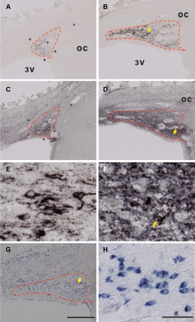
Fig. 2.
Immunocytochemical staining of arginine vasopressin (AVP), glutamic acid decarboxylase (GAD)65/67, and in situ hybridization (ISH) of GAD67-mRNA in the suprachiasmatic nucleus (SCN). a, b, e AVP immunoreactivity (ir), c, d, f GAD65/67-ir, and g, h GAD67-mRNA. C is an adjacent section to a, while d is an adjacent section to b, respectively. e, f, h Higher magnification of b, d, and g, respectively, from the area indicated by an arrow. Please note that AVP-staining is mainly present in neurons, but also in fibers, providing a clear boundary for the SCN (a, b, e), while GAD65/67-staining is largely present in SCN fibers (c, d, f) and only in a few of its neurons in some patients (indicated by an arrow in f). The SCN area was outlined according to an AVP-stained section (as shown by the dashed line), and the outline was transferred to the adjacent section with GAD65/67-staining or GAD67-mRNA ISH, based upon at least four markers in the area, as indicated by stars (usually blood vessels) in a and c. Please also note that almost all SCN neurons were surrounded by GAD65/67-ir beads (f), which probably represent synapses. In the rostral SCN, GAD-ir outlined the SCN (c) in almost the same way as AVP-ir (a), but in the more caudal part of the SCN, GAD65/67-staining was also distributed over a larger hypothalamic area around the SCN except for the optic tract and optic chiasm (OC) (d). GAD67-mRNA was abundantly distributed over the SCN and its surroundings (g). 3V third ventricle. Scale bar in a–d, g 0.5 mm, e, f, h 0.05 mm

