Figure 5.
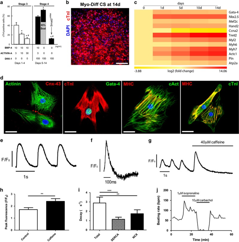
A stage-specific TGF-β-family/Wnt Inhibitor cocktail induces c-kitpos CSC CSs to differentiate with high efficiency into spontaneously rhythmic beating functional CMs in vitro. (a) Frequency of cTnI-positive CM-committed cells and percentage of beating cells resulting from the indicated different TGF-β/Wnt cocktail strategies. *P<0.05 versus all. #versus stage 4. Data are mean±S.D., n=6. (b) Representative confocal image shows cTnI (red) CM-committed cells at 14 days. DAPI stain nuclei in blue. Bar=50 μm. (c) qRT-PCR analysis heat map show sequential upregulation of cardiac transcription factors, followed by contractile genes with downregulation of cell cycle competence genes in differentiating c-kitpos CSC CSs at different time points in the stage-specific TGF-β-family/Wnt inhibitor cocktail. Colour scale indicates change in Ct (threshold cycle) relative to the normalised GAPDH control. (d) CSC-derived contracting CMs express contractile proteins (Actinin, cTnI, MHC and cardiac Actin) with co-expression of cardiac transcription factor (Gata-4). The CSC-derived CMs exhibit well-defined sarcomeric structures (z lines and dots) as well as gap junction formation (Cnx-43) between cells. DAPI stain nuclei in blue. Bar=20 μm. (e) Representative traces of calcium transients stimulated at 1 Hz, with change in fluo-4 fluorescence normalised to baseline (F/F0). (f) Representative trace of an action potential stimulated at 1 Hz, with change in di-8-anepps fluorescence normalised to baseline (F/F0). (g) Representative trace of a caffeine-induced calcium transient, which is preceded by three normal calcium transients stimulated at 1 Hz with change in fluo-4 fluorescence normalised to baseline (F/F0). (h) Peak fluorescence (F/F0) compared between control calcium transients and caffeine-induced calcium transients indicating a functional sarcoplasmic reticulum (**P<0.01, n=6). (i) Rate of calcium transient decay where kTotal=kSERCA+kNCX+kSlow with proportion of calcium extruded by each mechanism represented below (**P<0.01, ***P<0.005, n=6). (j) Example trace of change in spontaneous beating rate with application of the non-selective β-adrenergic agonist, isoprenaline and the muscarinic acetylcholine receptor agonist, carbachol

