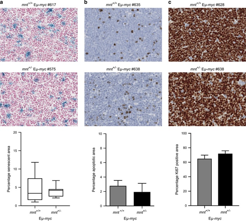
Figure 7.
Impact of mnt heterozygosity on Eμ-myc lymphoma cells. (a) mnt heterozygosity has no impact on senescence. Sections of lymph node lymphomas of mnt+/+ Eμ-myc (n=12) and mnt+/− Eμ-myc (n=7) mice were stained for β-galactosidase activity, counterstained with nuclear fast red (see Materials and Methods) and scanned on an Aperio Digital Pathology Slide Scanner. β-Galactosidase-positive cells in extracted images were quantified using Fiji software. Data shown are mean±S.E.M. (b) mnt heterozygosity does not impact upon apoptosis. Sections of lymphomas in lymph node and/or spleen of mnt+/+ Eμ-myc (n=10) and mnt+/− Eμ-myc mice (n=5) were stained using an antibody specific for cleaved caspase-3 followed by a haematoxylin counterstain (see Materials and Methods). Cells positive for cleaved caspase-3 were quantified as in (a) above. Data shown are mean±S.E.M. (c) Proliferation in Eμ-myc lymphomas is not altered by mnt heterozygosity. Sections of lymphomas in lymph node and/or spleen of mnt+/+ Eμ-myc (n=11) and mnt+/− Eμ-myc mice (n=4) were stained for Ki-67 with haematoxylin counterstain. The Ki-67 positive area was quantified as in (a) above. Data shown are mean±S.E.M.

