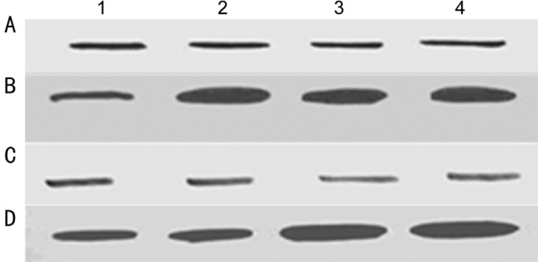
Figure 3. Western blot analysis of p53 (A) and Bcl-2 (B) in Y79 cells (lane 1) and Y79/vincristine cells (lane 2), Y79/etoposide cells (lane 3), and Y79/carboplatin cells (lane 4); Western blot analysis of p53 (C) and Bcl-2 (D) in primary cultures of primarily enucleated eyes (lanes 1&2) and in primary cultures of enucleated eyes after chemotherapy (lane 3&4).
Cell lysates were resolved by SDS-PAGE, and proteins were immunoblotted and detected using specific antibodies.

