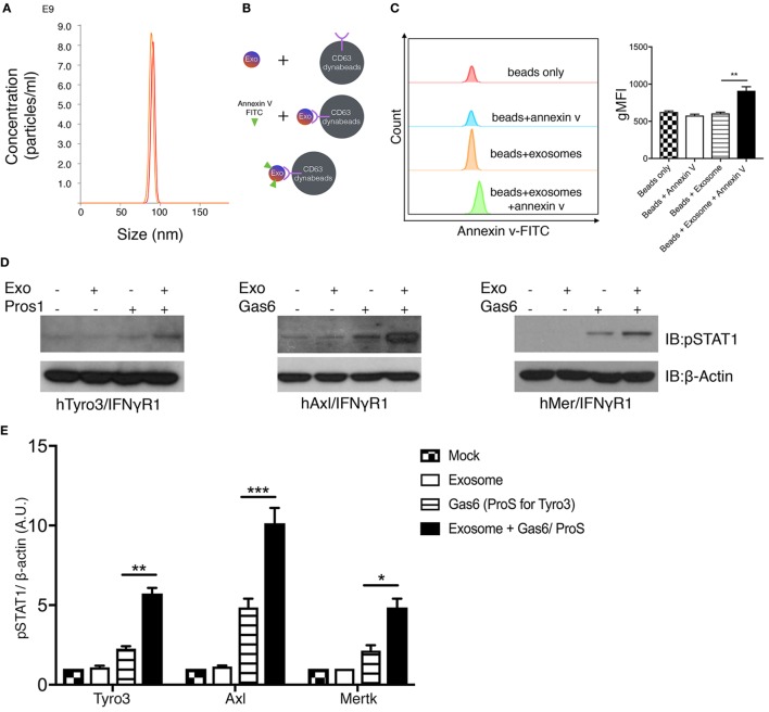
Figure 5.
Phosphatidylserine (PS)-positive tumor exosomes enhance Tyro3, Axl, and Mertk (TAMs) activation by growth arrest-specific 6 (Gas6) or protein S (Pros1). (A) Tumor-derived exosomes were purified from MDA-MB-231 cell’s culture media, and the size and concentration of the exosomes were analyzed by NanoSight Tracking Analysis. (B) Schematic procedure of PS staining of exosomes using CD63-Dynabeads. Exosomes isolated from MDA-MB231 cells were incubated with CD63-Dynabeads and then stained with FITC-Annexin V to assess PS exposure on the surface of exosomes. The experiment results are shown in (C) in which the samples from top to bottom are: beads alone (red), beads + FITC-Annexin V (blue), beads + exosomes (orange), beads + exosomes + FITC-Annexin V (green). The MFI quantification is shown in the right panel from independent experiments (n = 3). Error bar, SEM, **p < 0.005. (D) Exosomes that were isolated from MDA-MB231 cells were mixed with Gas6-containing media (Gas6-CM) or Pros1 (100 nM) to stimulate hTAM/IFNγR1 reporter cells. The activation of TAM receptors was assessed by pSTAT1 immunoblotting. (E) Densitometric quantification of hTAM/IFNγR1 reporter cells’ activation by exosomes (pSTAT1) normalized to β-actin from independent experiments (n = 3). Error bar, SEM. *p < 0.05, **p < 0.005, ***p < 0.001.

