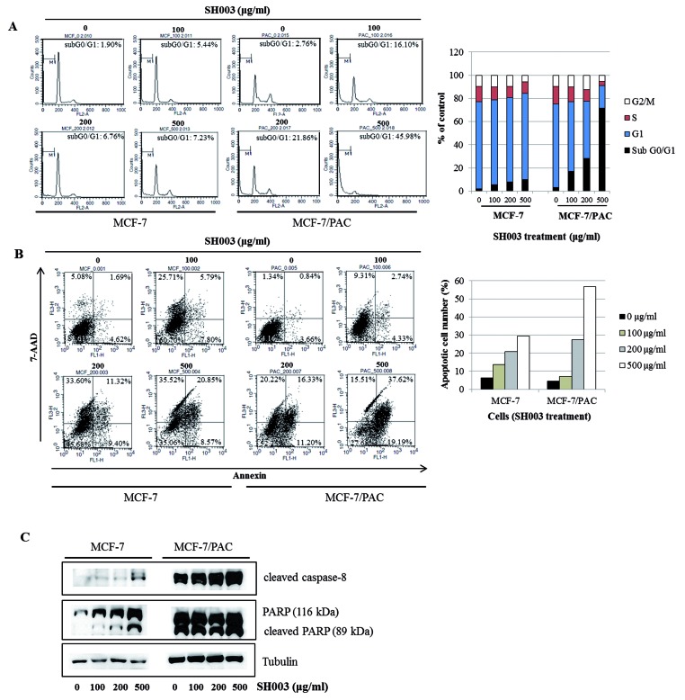
Figure 2. SH003 induces apoptosis in MCF-7/PAC cells.
(A) MCF-7 and MCF-7/PAC cells were treated with SH003 (0–500 μg/ml) and fixed 72 h later for flow cytometry. Propidium iodide-labeled nuclei were analyzed for DNA content. The sub-G0/G1 apoptotic population was quantified. (B) MCF-7 and MCF-7/PAC cells were treated with SH003 (0–500 μg/ml) and harvested after 72 h. Cells were stained with 7-AAD and annexin V-FITC. Apoptotic cell death was analyzed with a BD FACSCalibur flow cytometer using the FL1 and FL3 channels. (C) MCF-7 and MCF-7/PAC cells were treated with SH003 (0–500 μg/ml) for 24 h. Whole cell lysates were analyzed by Western blotting with anti-cleaved caspase-8, anti-PARP, and anti-tubulin antibodies. The data shown are representative of three independent experiments that gave similar results.

