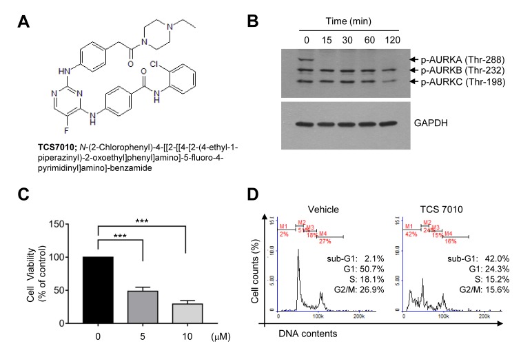
Figure 1.
Effect of TCS7010 on the induction of apoptosis. (A) Chemical structure of TCS7010. (B) HCT116 cells were treated with 5 µM TCS7010 for different periods of time (0–120 min). Whole cell lysates were analyzed using immunoblotting. GAPDH was used as an internal control. (C) Cell viability assay in which HCT116 cells were treated with 5 µM TCS7010 for 24 h. Cell viability was determined using a Cell Counting Kit-8 assay. Data are presented as the mean ± standard deviation. ***P<0.001 (n=3), one-way analysis of variance followed by Sidak's multiple comparisons test. (D) Cell cycle analysis in which HCT116 cells (1×10⁵ cells/sample) were treated with vehicle (DMSO) or 5 µM TCS7010, fixed with ethanol and stained with propidium iodide. The cellular DNA content was determined using flow cytometry. AURK, aurora kinase.

