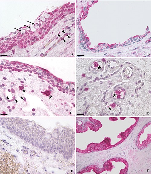
Figure 1.

Immunohistochemical staining of VDR. A,C,D,E) Pterygium; B) normal conjunctiva; F) human prostate. Pterygium showed a moderate to strong immunoreactivity of the epithelial cells, mostly localized in both nucleus and cytoplasm (A), while conjunctival epithelium exhibited mainly a mild to moderate cytoplasmic staining (B). In pterygium, the nuclear staining was always stronger than the cytoplasmic. A marked VDR immunoreaction was also detected in epithelial goblet cells (A) (arrows) and in the endothelial cells of sub-epithelial microvessels (A,C) (arrowheads). Scattered immunoreactive leucocytes-like cells, morphologically recognizable as cells belonging to the “mononuclear phagocyte system (MPS)”, were found in the subepithelial connective tissue (C) and inside the vessels (D) (asterisks). Negative (E) and positive (F) control sections. Scale bars: 20 μm.
