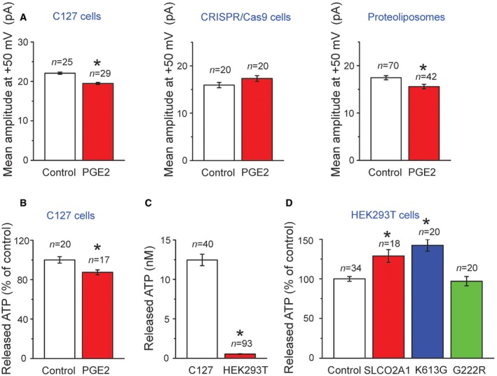
Averaged single‐channel amplitudes recorded at +50 mV in patches excised from C127 cells (left graph, n = 25 for Control and 29 for PGE2), from CRISPR/Cas9 KO C127 cells (center graph, n = 20 for Control and 20 for PGE2), and from giant proteoliposomes with reconstituted recombinant SLCO2A1 protein (right graph, n = 70 for Control and 42 for PGE2) in the absence (Control) and presence of 20 μM PGE2 added to bath and pipette solutions. The data from 4 to 9 cells and 4 proteoliposomes for each value are pooled.
Swelling‐induced ATP release from C127 cells in the absence (Control, n = 20) and presence of 20 μM PGE2 (n = 17). To test PGE2 effects, the cells were pre‐incubated with Ringer solution containing PGE2 and challenged with hypotonic medium containing PGE2.
Comparison between the absolute level of the swelling‐induced ATP release from (n = 40) and that from HEK293T cells (n = 93).
Swelling‐induced release of ATP from HEK293T cells transfected with mock (Control,
n =
34)), WT SLCO2A1(
n =
18) or with the mutants K613G (
n =
20) and G222R (
n =
20; see
Appendix Table S2 for the expression vectors used here).
Data information: Error bars, SEM. * indicates significantly different from Control C127 cells (A–C, by Student's
t‐test) or mock‐transfected Control HEK293T cells (D, by ANOVA) at
P <
0.05.