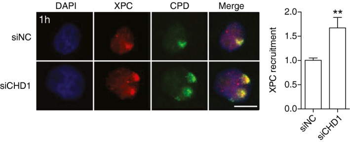
Figure EV3. XPC accumulation in the chromatin of UV‐irradiated HeLa cells.

Representative immunofluorescence images of HeLa cells that were UV‐irradiated (dose applied to filters: 100 J/m2) through micropore filters to generate local spots of DNA damage. Immunostaining was carried out after 1 h with antibodies against CPDs and XPC protein. Cells were pretreated 2 days earlier with siRNA targeting the CHD1 transcript (siCHD1) or with non‐coding control RNA (siNC). DAPI was used to stain nuclear DNA. Scale bar: 10 μm. The recruitment of XPC protein was quantified by measuring spot intensities followed by normalization to the nuclear background. Control values were set to 1. Data are presented as mean ± SEM (n = 3, 100 cells for each experiment). **P ≤ 0.01 (unpaired, two‐tailed t‐test).
