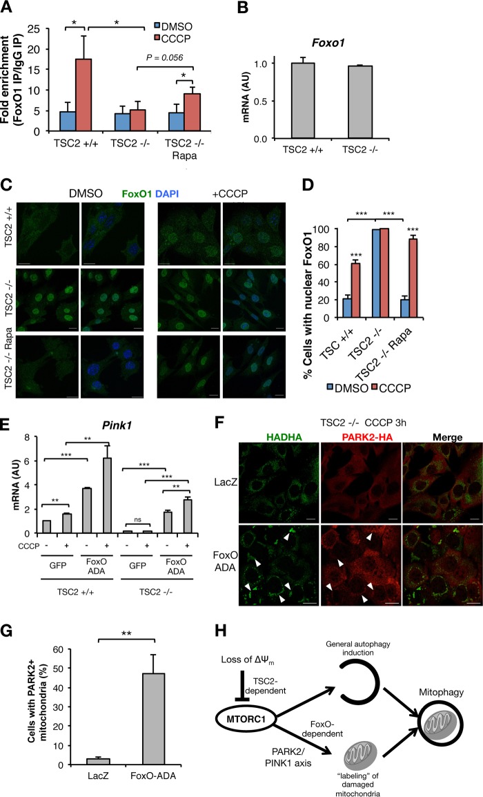
FIG 7.
FoxO-ADA rescues Pink1 expression and PARK2 translocation to mitochondria. (A) ChIP, using anti-FoxO1 or IgG control, in TSC2+/+ and TSC2−/− MEFs stimulated with CCCP or vehicle for 15 h with or without Rapa pretreatment, followed by qPCR for the Pink1 promoter region. (B) qPCR in TSC2+/+ and TSC2−/− MEFs. (C and D) Representative images of FoxO1 localization (C) and percentages of cells with nuclear FoxO1 (D) in TSC2+/+ and TSC2−/− MEFs stimulated with CCCP or vehicle for 15 h, with or without Rapa pretreatment. (E) qPCR in TSC2+/+ and TSC2−/− MEFs transduced with adenovirus encoding GFP or FoxO-ADA, with or without 15 h of exposure to CCCP. (F and G) Representative images of PARK2 mitochondrial localization (arrowheads) (F) and percentages of cells with PARK2+ mitochondria (G) in TSC2+/+ and TSC2−/− MEFs stably expressing PARK2-HA and transduced with LacZ or FoxO-ADA and then treated for 3 h with CCCP. The values represent means and SD (n = 3 to 5 independent experiments). *, P < 0.05; **, P < 0.01; ***, P < 0.001; ns, not significant compared to the indicated control. (H) Model in which loss of ΔΨm leads to TSC2-dependent MTORC1 inhibition, fundamental to coordinating two synergistic MTORC1-dependent processes—autophagy initiation and labeling of the cargo to be degraded, enabling its recognition by the autophagic machinery—to execute the clearance of damaged mitochondria.

