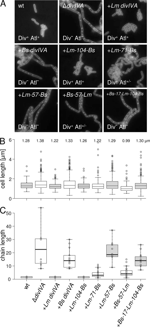
FIG 2.
Effect of chimeric DivIVA proteins on cell length and chaining of L. monocytogenes. (A) Fluorescence micrographs showing Nile red-stained cells. Strains EGD-e (wt), LMS2 (ΔdivIVA), LMS30 (+Lm divIVA), LMKK8 (+Bs divIVA), LMKK13 (+Lm-104-Bs divIVA), LMS157 (+Lm-71-Bs divIVA), LMKK12 (+Lm-57-Bs divIVA), LMKK14 (+Bs-57-Lm divIVA), and LMS149 (+Bs-17-Lm-104-Bs divIVA) were grown in BHI broth containing 1 mM IPTG at 37°C to mid-logarithmic growth phase and analyzed by microscopy. Div and Atl phenotypes are indicated. (B) Cell lengths of 300 cells per strain were measured, and the length distribution is illustrated in a box plot (http://shiny.chemgrid.org/boxplotr/). Center lines show the medians; box limits indicate the 25th and 75th percentiles, as determined by the R software; whiskers extend 1.5 times the interquartile range from the 25th and 75th percentiles; and outliers are represented by dots. n = 300 sample points. Median cell lengths are given above the diagram. (C) Lengths of cell chains of ∼200 cells per strain were measured and illustrated in the same kind of box plot as in panel B.

