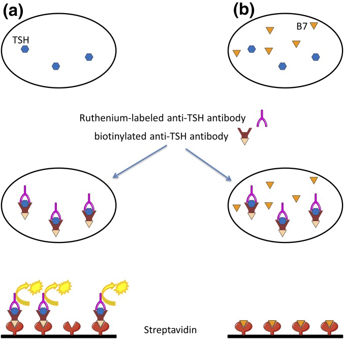
Figure 1.
Mechanisms of biotin interference in the TSH assay. (a) The serum sample is incubated with a biotinylated monoclonal anti-TSH antibody and a ruthenium-labeled monoclonal anti-TSH antibody. The addition of streptavidin-coated magnetic microparticles causes the resulting immune complexes to bind to the solid phase. Application of a voltage generates chemiluminescence, in direct proportion to the TSH level. (b) A high biotin concentration in the sample saturates streptavidin binding sites, yielding a falsely low TSH level. B7, biotin.

