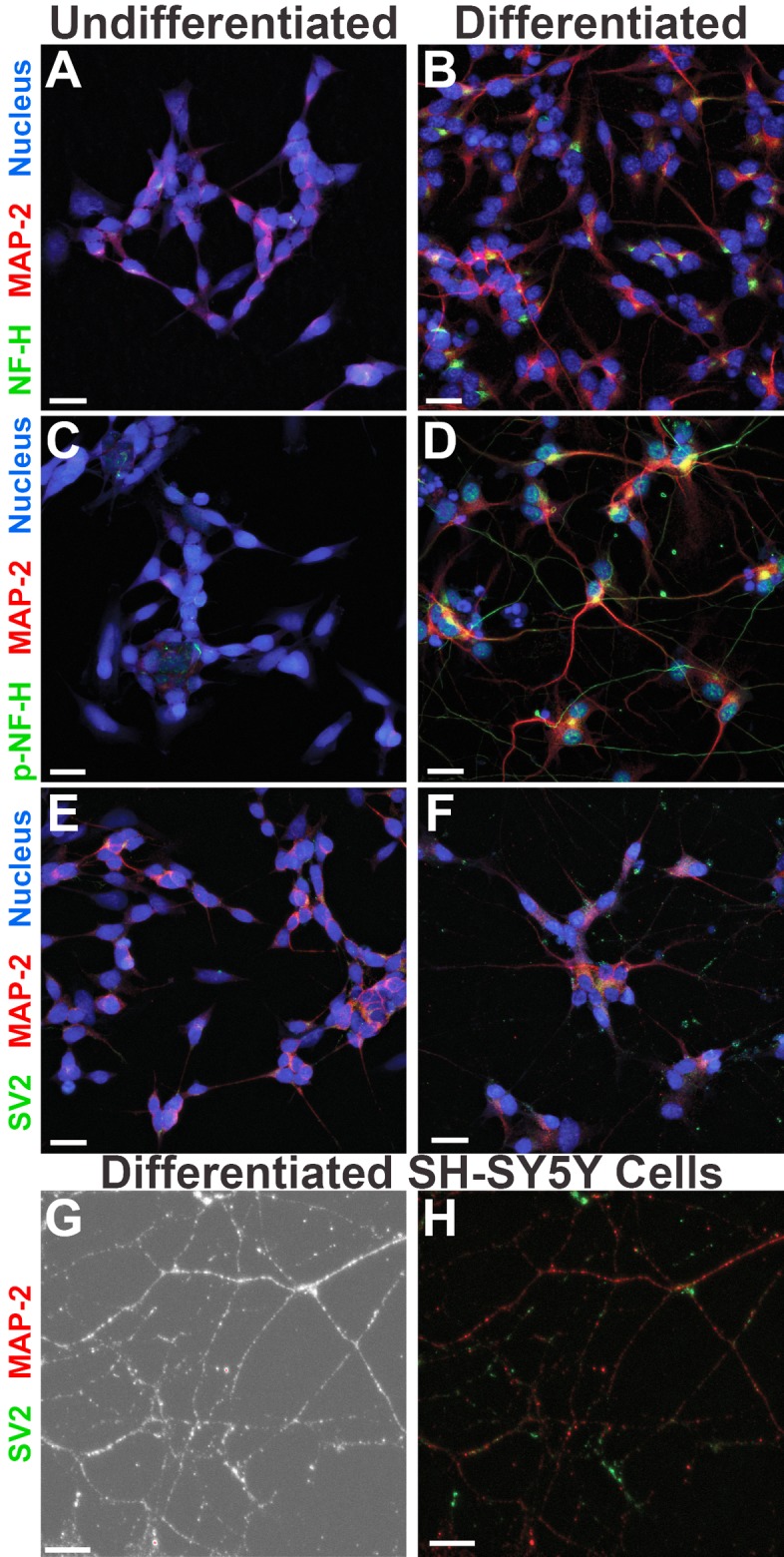
FIG 2.

Terminally differentiated SH-SY5Y neuronal cells show distinct localization of cytoskeletal and synaptic markers. Differentiation of SH-SY5Y cells for 2.5 weeks yields morphologically distinct localization of neuronal proteins (B, D, and F), in contrast to that seen in undifferentiated SH-SY5Y cells (A, C, and E). Higher magnification of differentiated SH-SY5Y neurites shown in phase-contrast (G) reveals further details of microtubule and synaptic vesicle staining (H). The 2.5-week differentiation process includes the addition of RA, neurotrophic factors, and ECM proteins. MAP-2, microtubule associated protein 2; NF-H, unphosphorylated neurofilament heavy chain; p-NF-H, phosphorylated neurofilament heavy chain; SV2, synaptic vesicle protein 2; Nucleus, nuclear/DNA stain. Images were taken with an Olympus FV10i confocal microscope at a magnification of ×60, and the scale bars represent 20 μm. Images in panels G and H were taken with an additional 2.6× digital zoom, and the scale bars represent 10 μm.
