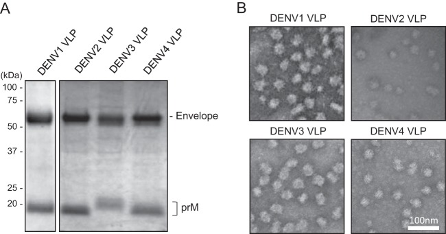
FIG 5.
Characterization of DENV VLPs. (A) SDS-PAGE analysis of purified DENV1 to -4 VLPs. Separated proteins were stained with Coomassie blue dye. Images representative of at least 3 replicates are shown. (B) Transmission electron microscopy images of purified DENV1 to -4 VLPs. Representative images are shown.

