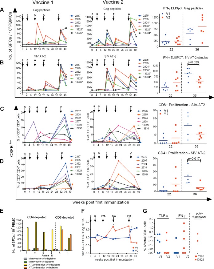
FIG 6.
Cellular immune responses throughout immunization. (A and B) IFN-γ ELISpot responses in blood of vaccinees during immunization are presented as SFCs per 106 PBMCs after stimulation with a SIVGag 15-mer peptide pool (A) and SIV AT-2 (B). Individual values are shown. The four- and five-digit numbers are monkey designations. The arrows indicate immunization with either SCIV (S), adenovirus (A), or fowlpox virus (F). The asterisks mark monkeys with MHC class I genotype Mamu-A1*001:01. (Right) Values for week 22 and week 36 after initial immunization are shown for group comparison. (C and D) Proliferative CD8+ and CD4+ T-cell responses in blood of vaccinees against SIV AT-2 determined by a CFSE proliferation assay shown as individual percentages of responding CD8+ (C) and CD4+ (D) T cells after initial immunization. Monkeys are indicated by four- or five-digit numbers. (C and D, right) Comparisons between values for week 22 and week 36 after initial immunization. At week 36, data from only 5 animals were available for V1. (E) Cell-mediated responses measured by ELISpot assay upon SIV AT-2 stimulation are CD4+ T-cell restricted. To identify which T-cell subsets were activated through SIV AT-2 stimulation in the IFN-γ ELISpot assay, we used PBMCs from long-term SIV-infected macaques known to be strongly reactive against SIV AT-2. IFN-γ ELISpots were performed with PBMCs without depletion (SIV AT-2 stimulation w/o depletion) and after depletion (SIV AT-2 stimulation w depletion) of CD4+ (n = 4) and CD8+ (n = 2) cells and the corresponding negative controls (microvesicle w/o depletion and microvesicle w depletion). (F) The ratio of the ELISpot results after stimulation with SIV AT-2 and Gag peptides was calculated for each individual monkey, and the kinetics of the ratio (median) of the two vaccine groups are displayed. The asterisks indicate significance between the two vaccine groups (Mann-Whitney test; P < 0.05). (G) Percentages of Gag-specific CD8+ T cells among total CD8+ CD3+ cells expressing either TNF-α or IFN-γ determined by ICS at week 36 in V1 (blue) and V2 (red). Polyfunctional CD8+ cells expressing IFN-γ plus IL-2, IFN-γ plus TNF-α, and IFN-γ plus IL-2 plus TNF-α observed only in animal 2295 (V2) are shown cumulatively. IFN-γ- plus TNF-α-expressing cells were detected in animal 13929 (V2).

