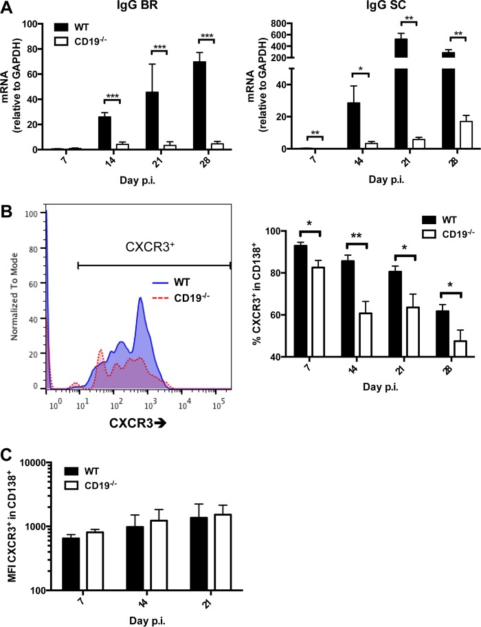
FIG 6.
Total CXCR3+ ASC are reduced in CLN of CD19−/− mice. (A) Brains (BR) and spinal cords (SC) harvested from infected WT and CD19−/− mice at the indicated times p.i. were analyzed for expression of IgG heavy chain (Ighg) mRNA. The data represent the means and SEM of transcript levels relative to gapdh mRNA of individual mice from 2 separate experiments, each comprising 2 to 6 individual mice per time point and group. (B) CLN cells pooled from infected mice were stained for CD138 and CXCR3. Representative histograms gated on B220+ CD138+ ASC at 21 days p.i. are shown for WT and CD19−/− mice; the graph depicts mean and SEM percentages of CXCR3+ cells within ASC (n = 2 to 4 individual mice per group per time point from 3 separate experiments). Statistically significant differences between WT and CD19−/− mice are denoted by asterisks: *, P < 0.05; **, P < 0.01; ***, P < 0.001. (C) MFI of CXCR3+ ASC from the cells depicted in panel B.

