
FIG 5.
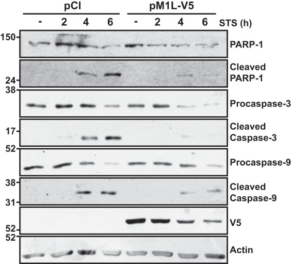
Biochemical hallmarks of staurosporine-induced apoptosis are reduced when M1 is expressed independently of infection. Subconfluent HeLa cellular monolayers were transfected with 1 μg of either empty vector (pCI) or pM1L-V5. At 24 h posttransfection, cells were treated with medium either lacking (−) or containing 0.5 μM staurosporine (STS). At 2, 4, or 6 h post-STS incubation, cells were collected and lysed in RIPA buffer. Lysates were separated by SDS–12% PAGE, and proteins were transferred to a PVDF membrane. Membranes were probed with either anti-PARP-1, anti-caspase-3, or anti-caspase-9 antisera. Membranes were developed and bands were detected using chemiluminescence. Blots were subsequently incubated with either anti-V5, anti-FLAG, or anti-actin antiserum and redeveloped. Data shown are representative of at least three independent experiments.