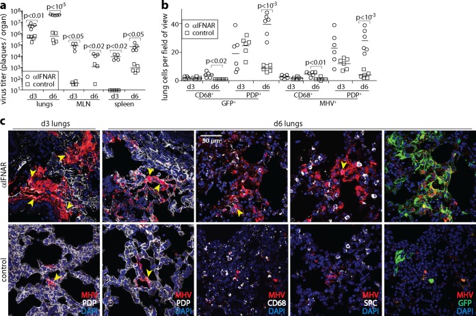
FIG 1.
IFNAR blockade increases MuHV-4 lytic infection of AM and AEC1 in the lungs. (a) C57BL/6 mice were given an IFNAR-blocking antibody i.p. (αIFNAR) or not (control) and then given MHV-GFP i.n. (104 PFU in 30 μl under anesthesia). At days 3 and 6, the titers of infectious virus in lungs were determined by a plaque assay, and the titers of total recoverable virus (lytic plus latent) in MLN and spleens were determined by an infectious-center assay. Symbols show data for individual mice. Bars show means. Significant differences are indicated. (b) After infection as described above for panel a, day 3 and day 6 lung sections were stained for virus-expressed GFP and lytic antigens (MHV). Lung myeloid cells, predominantly AM, were identified by staining for CD68. AEC1 were identified by staining for podoplanin (PDP). We counted infected cells in at least 4 fields of view per section, across 2 sections from each of 3 mice per group. Symbols show mean counts per section. Bars show group means. Significant differences between groups are shown. (c) In representative images of MHV staining quantitated as described above for panel b, yellow arrows show example dual-positive cells. Colocalization with surfactant protein C-positive (SPC+) AEC2 was rare (<2% of infected cells). Dual staining is also shown for GFP and MHV (colocalization appears in yellow). Viral GFP expression was generally more extensive than MHV staining, implying that not all infection was lytic, but it had a similar distribution.

