FIG 3.
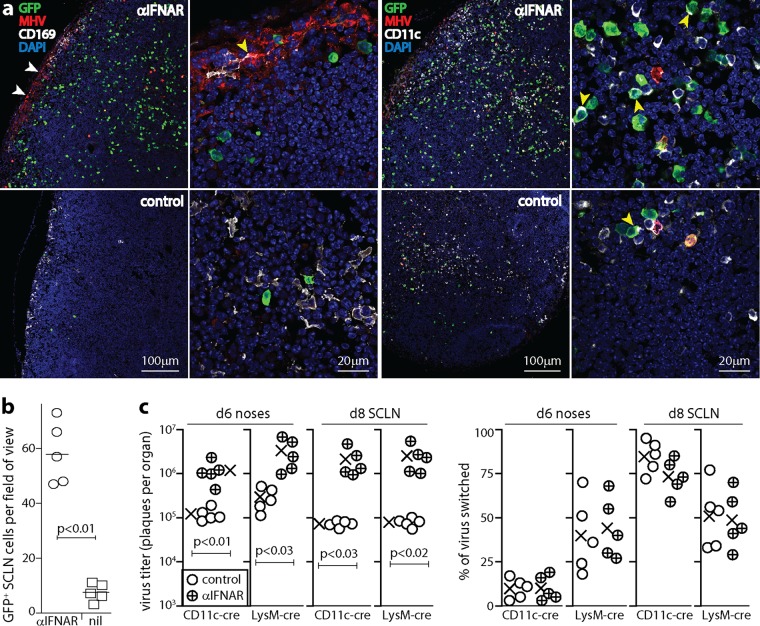
IFNAR blockade increases MuHV-4 LN infection. (a) C57BL/6 mice were given anti-IFNAR treatment or not (control) and then given MHV-GFP i.n. (105 PFU in 5 μl). At day 6, SCLN sections were stained for viral GFP and lytic antigens (MHV), for CD169 to identify SSM, and for CD11c to identify DC. Nuclei were stained with DAPI. Low-magnification images (left panels of each antibody set) show marked increases of MHV staining around the subcapsular sinus (white arrows) and GFP staining throughout the LN with anti-IFNAR treatment. GFP is expressed from an EF1α promoter in the viral genome, independently of lytic infection (55). CD11c staining of SSM is weak and thus is difficult to see at a low magnification. High-magnification images (right panels of each antibody set) show the subcapsular sinus for CD169 (the yellow arrow shows an MHV+ SSM) and example GFP+ DC in the LN substance for CD11c (yellow arrows). (b) Quantitation of GFP+ cells across at least 2 SCLN sections for each of 3 mice per group shows a significant increase after anti-IFNAR treatment. Symbols show mean counts for each section (at least 5 fields of view per section). Bars show group means. (c) CD11c-cre and lysM-cre mice were treated or not with anti-IFNAR and then given MuHV-4 with a floxed color-switching cassette (MHV-RG) (105 PFU i.n. in 5 μl). Noses were assayed for infectious virus at day 6 by a plaque assay, and SCLN were explanted to recover both lytic and latent virus at day 8. Recovered plaques were also typed as being red (mCherry+, native) or green (GFP+, switched) fluorescent. Percent switching was calculated as follows: 100 × green plaques/(green plaques + red plaques). Circles show data for individual mice. Crosses show means. Significant differences are shown. Regardless of anti-IFNAR treatment, lysM-cre mice had more virus switching in noses, and CD11c-cre mice had more virus switching in LN (P < 0.02).

