FIG 7.
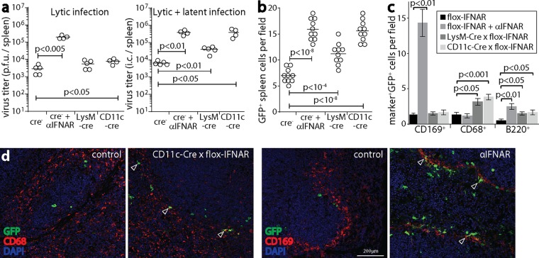
IFNAR signaling to myeloid cells restricts splenic infection by i.p. inoculation of MuHV-4. (a) CD11c-cre × flox-IFNAR, lysM-cre × flox-IFNAR, and cre− flox-IFNAR mice were given MHV-GFP i.p. (105 PFU). As a positive control, cre− mice were also given anti-IFNAR treatment. At day 4, spleens were assayed for lytic infection by a plaque assay and assayed for total recoverable virus (lytic plus latent infection) by an infectious-center (i.c.) assay. Circles show titers in individual mice. Bars show means. Significant differences are shown. (b) Mice were infected as described above for panel a. At day 4, spleen sections were stained for viral GFP. GFP+ cell counts across 10 sections from 3 mice for each group were increased in all groups compared to those for the cre− negative controls. Symbols show mean counts per section from at least 5 fields of view per section. Bars show group means. (c) Spleen sections, as described above for panel b, were stained for viral GFP plus markers of MZM (CD169), myeloid cells in general (CD68), and B cells (B220). Anti-IFNAR treatment increased MZM infection, whereas CD11c-cre and lysM-cre expression increased myeloid cell infection without a focus on MZM. All groups showed significantly more B cell infection than that in cre− negative controls. Bars show mean counts ± standard errors of the means for 10 sections from 3 mice, counting at least 5 fields of view per section. (d) Examples of myeloid cell staining from panel c show increased CD169+ cell infection with anti-IFNAR treatment and increased CD68+ cell infection with CD11c-cre expression. CD11c-cre+ and lysM-cre+ mice had infection around the MZ, but little infection colocalized with CD169, and GFP+ cells were more evenly distributed across the MZ and white pulp. Arrows show example positive cells.

