FIG 2.

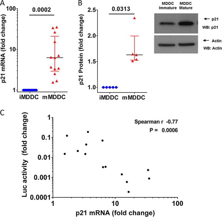
p21 induction in mature MDDCs correlates with viral replication levels. (A) p21 mRNA expression was quantified by qRT-PCR in immature (blue) and mature (red) MDDCs. The data are expressed as the fold change in the number of p21 copies in mature MDDCs compared to that in immature MDDCs. Each symbol represents the results obtained with cells from one donor (n = 13). The median and IQR values are shown. (B) Analysis of protein expression in immature and mature MDDCs by Western blotting (WB). (Left) p21 protein expression quantification are expressed as the relative change in p21 protein expression in mature MDDCs compared with that in immature MDDCs in the same experiments. The median and IQR values of independent experiments (n = 5 donors) are depicted. An example of p21 protein analyzed by Western blotting is shown. (C) Correlation between relative changes in the number of p21 mRNA copies and relative changes in luciferase activity after infection with HIV-1 NL4.3ΔenvLuc VSV-G in mature MDDCs compared with those in immature MDDCs from 14 different donors.
