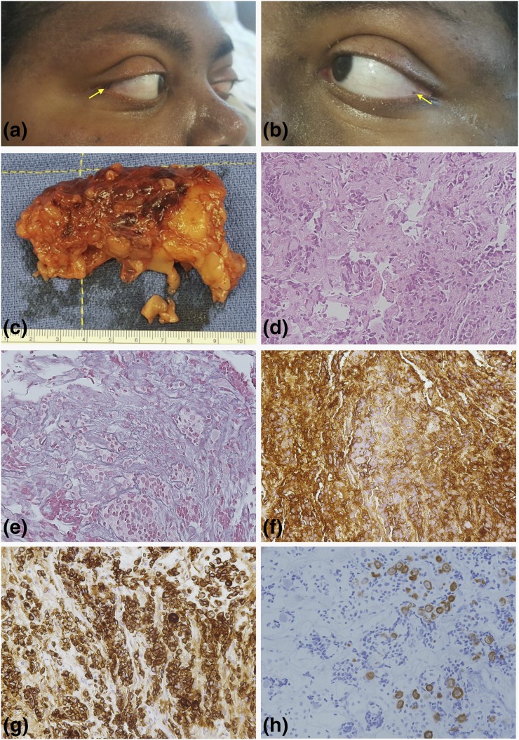
Figure 1.
Clinical and histopathological presentation. (a and b) Small epicanthal lentigines were observed in this patient. (c) The surgical specimens of bilateral adrenalectomy displayed the characteristics of PPNAD, and such diagnosis was later confirmed by histopathological examination. (d) Hematoxylin–eosin staining (20×) of the corticotropinoma tissue. The tumor was a microadenoma measuring approximately 6 × 4 × 2 mm, with Crooke’s cells surrounding the neoplastic tissue. (e) Breakdown of the reticulin network (20×), (f) as well as strong and diffusely positive ACTH staining (20×), was demonstrated. (g) Extensive positive immunostaining for CAM5.2 was identified (20×). (h) Keratin 20 immunostaining was found in some areas containing Crooke’s cells (20×). These images were compatible with a diagnosis of Crooke’s cell adenoma.

