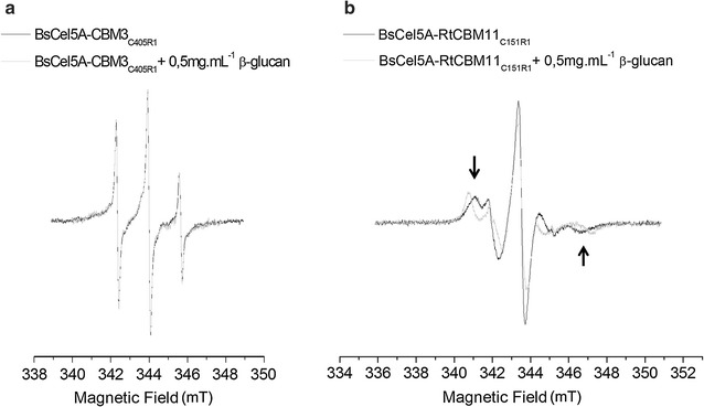
Fig. 3.

Measured EPR spectra of (a) BsCel5A-CBM3C405R1 and (b) BsCel5A-RtCBM11Y151R1 both free in solution and in the presence of β-glucan. The spectra are superimposed and normalized to the total area to emphasize the difference in line width and shape. The arrows highlight the increase in the out-peak separation of the spectrum due to β-glucan binding and immobilization of the spin-labeled side chain
