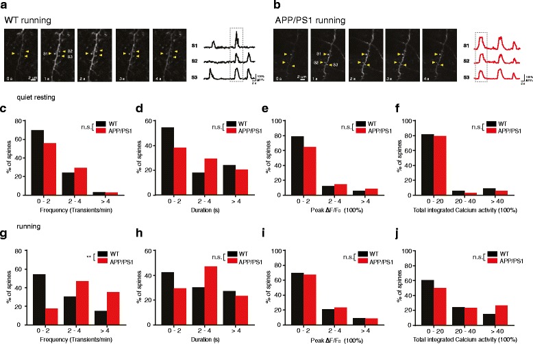
Fig. 2.

The overall synaptic activity on apical dendrites is comparable between WT and APP/PS1 mice. a, b. Time-lapse images of dendritic spine calcium transients (left) and fluorescent traces of dendritic spines expressing GCaMP6s (right) in WT (a) and APP/PS1 mice (b). Calcium transients in three spines (arrowheads point to each spine) were shown. c-f. Distributions of the frequency (c), duration (d), peak ΔF/F0 (e) and total integrated activity (f) of spine calcium transients in WT and APP/PS1 mice under quiet resting state (WT:4 mice, 32 spines; APP/PS1: 5 mice, 30 spines. Mann-Whitney U Test). g-j. Distributions of the frequency (g), duration (h),peak ΔF/F0 (i) and total integrated activity (j) of spine calcium transientsin WT and APP/PS1 mice during treadmill running (WT: 4 mice, 33 spines; APP/PS1: 5 mice, 34 spines. Mann-Whitney U Test). **P < 0.01; n.s., not significant
