Fig. 1.

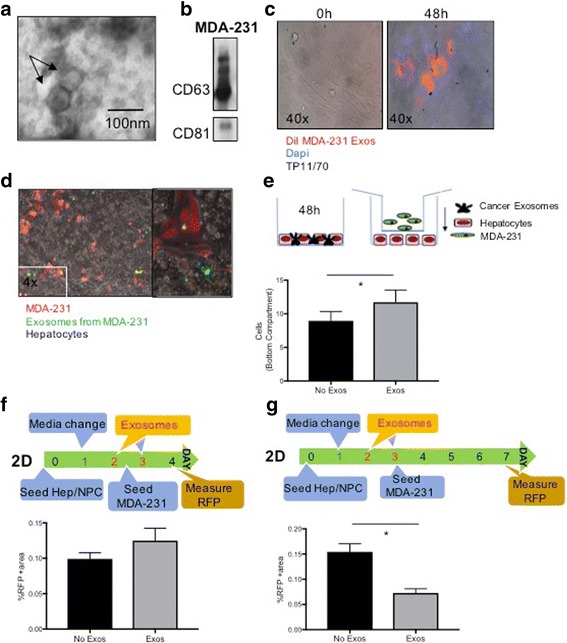
Breast cancer cell produce exosomes that interact with and alter human hepatocytes. a Transmission electron microscopy of concentrated exosomes in the media of MDA-231 cells. b Immunoblot analyses of the exosome-specific markers CD81, CD63 in MDA-231 exosome protein extracts. c MDA-231-derived exosomes were stained with DiI and overlaid on top of normal human fibroblasts TP11/70. The cells were then washed and fluorescence in the cells was assessed at 0 and 48 h. After fixing the cells at 48 h, DAPI was used for nuclear staining. d MDA-231 cells expressing RFP were transfected with CD63-GFP. These cells were cocultured with human hepatocytes, with the GFP showing uptake of exosomes distant from the RFP+ MDA-231 cells. Right panel shows higher magnification. e MDA-231 cells migrate towards exosome-primed hepatocytes. Human hepatocytes were seeded on the bottom of a transwell plate and primed with MDA-231-derived exosomes for 48 h. RFP+ MDA-231 cells were then added in the transwell insert (8um pores) and cocultured for another 48 h. Top: schematic of experiment. Bottom: quantitation of RFP+ cells among the human hepatocytes. f Primary human liver cells were plated for two days, followed by priming with MDA-231-derived exosomes on days 2 and 3. At day 3, RFP+ MDA-231 cells were added to the culture, and 24 h later, the intercalated cells enumerated. Top: schematic of experiment. Bottom: quantitation of RFP+ cells among the liver cells. g The experiment in F was evaluated four days after seeding the MDA-231 cells, at the time the primary human liver cells start to die. Top: schematic of experiment. Bottom: quantitation of RFP+ cells. A-D are representative examples of experiments performed at least 3 times, and E-G are mean ± s.e.m. of three experiments each performed in duplicate. * P < 0.05
