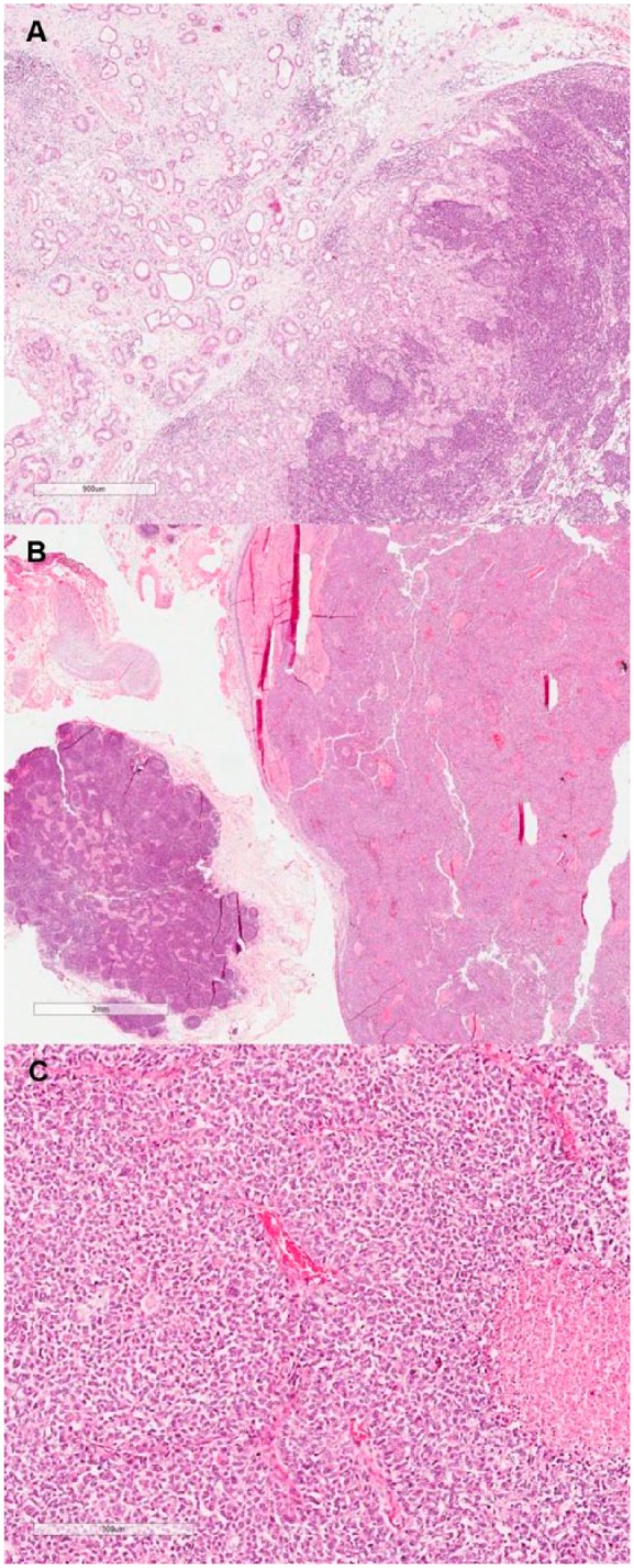
Figure 2.

Lymph node metastasis of the adenocarcinoma component (A; hematoxylin-eosin [HE], original magnification 100×) in contrast to another lymph node infiltrated by the neuroendocrine component (B; HE, original magnification 100×). High-power view of the neuroendocrine carcinoma metastatic component (C; HE, original magnification 200×).
