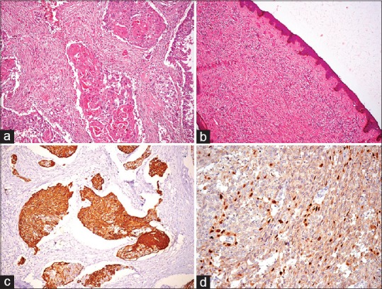
Figure 1.

(a) Infiltrating neoplasm composed of sheets and nests of atypical cells exhibiting squamous differentiation (H and E, ×200). (b) Section from nipple showing wavy spindle-shaped cells admixed with fibroblasts (H and E, ×200). (c) Immunohistochemistry for CK5/6 showing diffuse cytoplasmic positivity (×200). (d) Immunohistochemistry for P63 showing nuclear positivity (×200)
