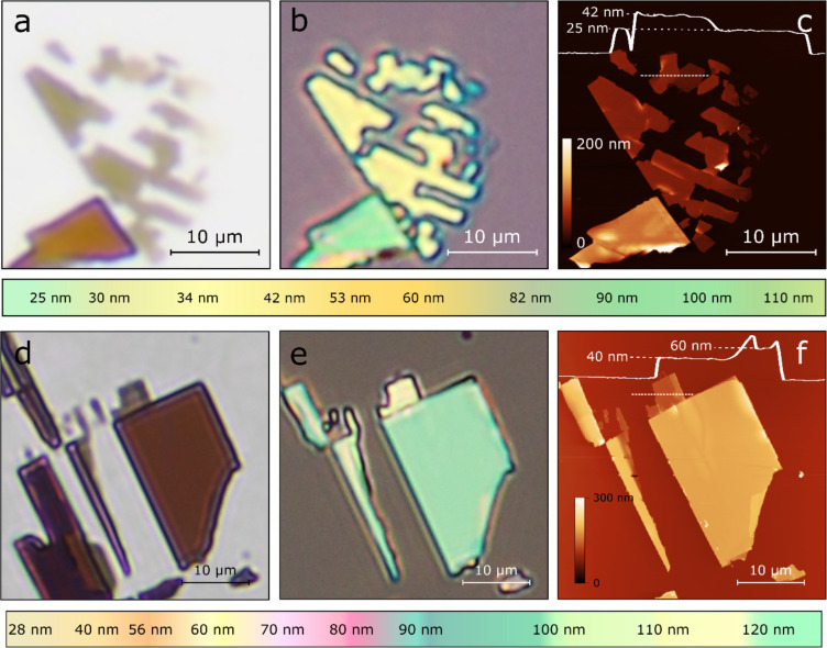
Figure 2.
(a) Transmission-mode optical microscopy image of franckeite flakes on a Gelfilm carrier substrate. (b) Epi-illumination optical microscopy image of the same franckeite flake after being transferred onto a 292 nm SiO2/Si substrate. (c) Atomic force microscopy image of the same flake to determine its thickness. Below (a) to (c) the colour chart shows a coarse guide to determine the thickness of franckeite flakes on 292 nm SiO2/Si substrates through their apparent colour. (d–f) Similar as (a) to (c) but for a franckeite flake transferred onto a 92 nm SiO2/Si substrate. Below (d) to (f) the colour chart shows a coarse guide to determine the thickness of franckeite flakes on 92 nm SiO2/Si substrates through their apparent colour.

