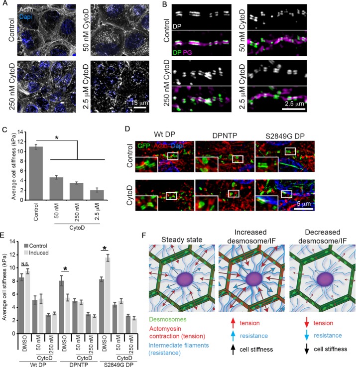
FIGURE 5:
The DSM–IF-mediated effects on cell stiffness are dependent on the actin cytoskeleton. (A) Apotome micrographs of cells treated with the indicated concentrations of the actin depolymerization agent CytoD are shown with actin filaments (phalloidin staining) in white and DAPI in blue to show nuclei. DMSO was used as the control. (B) Superresolution micrographs of cells treated with the indicated concentrations of CytoD are shown with staining of DP in white (above) and in green (below) overlaid with plakoglobin (PG) in magenta at representative cell–cell junctions. DMSO was used as the control. (C) Average cell stiffness measurements of individual cells within semiconfluent (80%) cell sheets for cells treated with the indicated concentrations of CytoD are shown. DMSO was used as a control. Error bars represent the standard error of the mean from at least 99 cells from three independent experiments. *, p < 0.0001. (D) Superresolution micrographs of induced A431 cells expressing GFP-tagged DP variants and treated with 250 nM CytoD are shown. The GFP-tagged DP variants are shown in green, actin filaments (phalloidin staining) are shown in white, and DAPI indicates nuclei in blue. (E) Average cell stiffness measurements of individual induced or uninduced (control) A431 cells within semiconfluent (80%) cell sheets and treated with the indicated concentrations of CytoD or DMSO as a control are shown. Error bars represent the standard error of the mean from at least 30 cells from three independent experiments. *, p < 0.01; N.S., not significant. (F) Model: the DSM–IF linkage regulates the balance of cell forces. Cells exist in a “prestressed” state, which allows the system to respond rapidly to mechanical stimuli. There are tensile (red) and compressive (blue) elements that resist this tension. The tensile component is widely thought to be composed of the actomyosin machinery. Our data support a model in which the DSM–IF network may be functioning to resist the tension generated by actomyosin contractility. In this model, strengthening the DSM–IF connection leads to an increased resistive capacity of the system, allowing for more robust actomyosin-generated tension and increased cell forces/stiffness. However, uncoupling the DSM–IF network would decrease the resistive capacity and lead to a decrease in actomyosin-generated tension and cell forces/stiffness.

