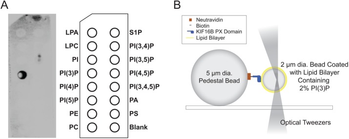
FIGURE 1:
(A) PIP-Strip showing the binding of biotinylated KIF16B-PX to PI(3)P as detected by chemiluminescence imaging of HRP-streptavidin. (B) Diagram of experimental setup used to measure the interaction of pedestal attached KIF16B-PX with membrane-coated beads held in an optical trap. The trap position was oscillated, resulting in the compression of the trapped beads against KIF16B-PX–coated pedestals, followed by retraction. Adhesions forces displace the bead from the trap center during retraction. Formation and subsequent rupture of bonds appeared as negative peaks in the data traces (Figure 2A). Beads and protein molecules are not drawn to scale. Adapted from Pyrpassopoulos et al. (2013).

