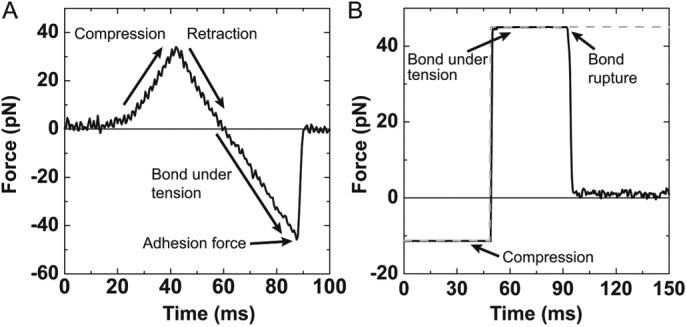
FIGURE 2:

(A) Example of an experimental ramp-force rupture event between 2% PI(3)P lipid-coated bead and KIF16B-PX at a loading rate of 1500 pN/s. The three regions are the compression of the optically trapped bead on the pedestal, retraction of the bead in the opposite direction until the compressive force reaches zero, and the bond under tension as the optical trap is moved until the bond ruptures. The reported adhesion force is the point where the bond ruptures. (B) The attachment duration of an experimental single adhesive bond between a 2% PI(3)P lipid-coated bead and KIF16B-PX under 45 pN of load. A square pulse (dashed gray trace) is the signal to drive compression and retraction of the membrane-coated bead. Adhesive forces (solid black trace) are recorded during attachments. A constant force is maintained via a feedback loop until the bond ruptures.
