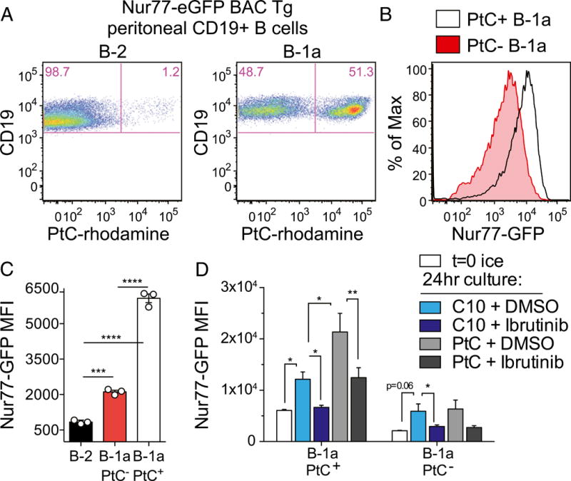
FIGURE 2. Nur77-eGFP expression identifies self-reactive PtC-specific B-1a cells in vivo.

(A) PerC cells from Nur77-eGFP reporter mice were stained immediately ex vivo with surface markers and PtC-rhodamine liposomes to identify B cell subsets (as gated in Fig. 1A). Representative plots show PtC+ B cells in the B-2 and B-1a compartments of these mice. (B) Line graph depicts Nur77-eGFP expression in PtC+ and PtC− B-1a cells gated as in (A). (C) Graph depicts Nur77-eGFP mean fluorescence intensity (MFI) (± SEM) in PerC B cell subsets stained and collected by flow immediately ex vivo and gated as in (A). ***p < 0.0005, ****p < 0.0001, unpaired t test. (D) PerC cells from Nur77-eGFP reporter mice were incubated in complete culture media for 24 h with ibrutinib or DMSO in the presence or absence of PtC-rhodamine liposomes. Cells were then stained, collected, and analyzed as in (A). Bar graph depicts GFP MFI (± SEM) in PtC+ and PtC− B-1a cells in n = 3 biological replicates. *p < 0.05, **p < 0.005, ratio paired t test.
