Figure 1.
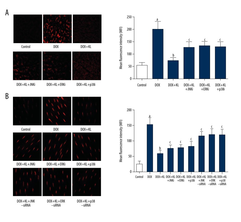
Klotho reduced DOX-induced ROS in myocardium and myocytes which was reversed by MAPKs inhibitors or siRNAs. (A) Left panel demonstrates the captured fluorescent images of DHE stained myocardium. Intracellular ROS is tagged red. Columns on the right side indicate the mean fluorescence intensities of ROS in myocardium harvested from control, DOX, DOX+KL, DOX+KL+JNKi, DOX+KL+ERKi and DOX+KL+p38i respectively. (B) Images on the left side are DHE staining of cultured myocytes. Columns on the right side indicate the mean fluorescence intensities of ROS in myocardium harvested from control, DOX, DOX+KL, DOX+KL+JNKi, DOX+KL+ERKi, DOX+KL+p38i, DOX+KL+JNK-siRNA, DOX+KL+ERK-siRNA, and DOX+KL+p38-siRNA respectively. a Differences were significant when compared with control (p<0.05); b differences were significant when compared with DOX (p<0.05); c differences were significant when compared with DOX+KL (p<0.05).

