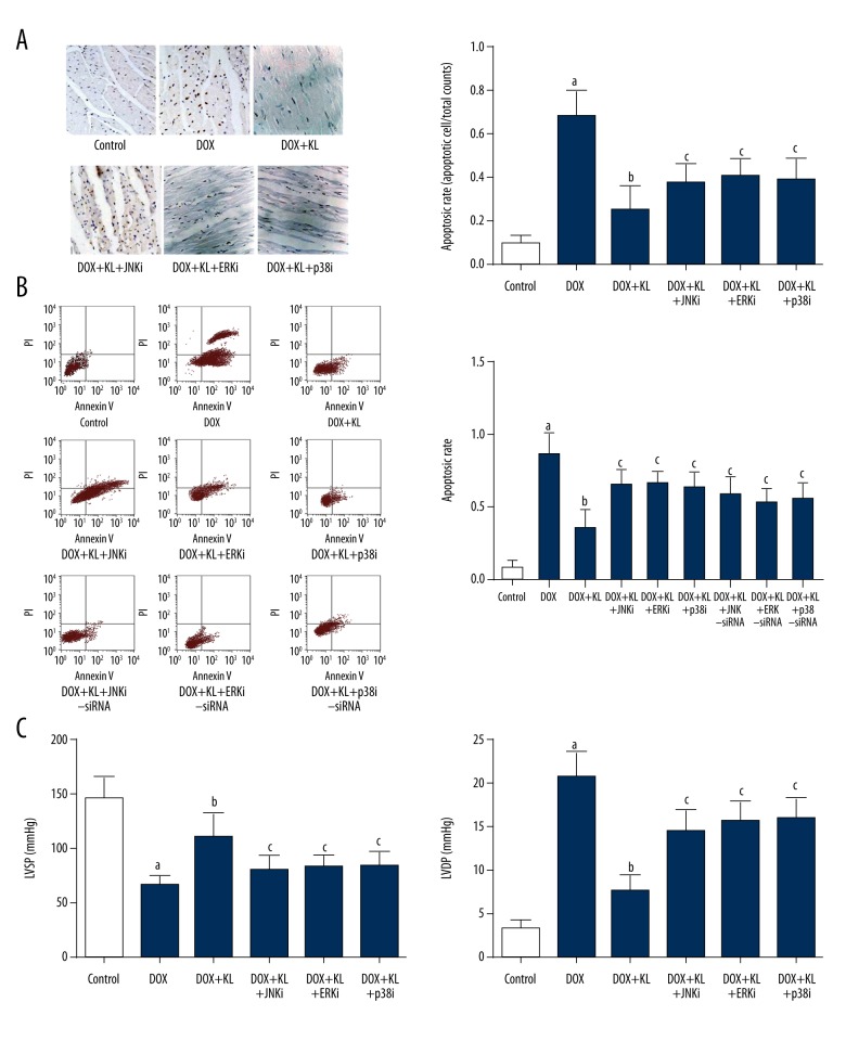
Figure 5.
Klotho attenuated DOX-induced apoptosis and cardiac dysfunction which were reversed by treatment of MAPKs and siRNAs. (A) Left panel demonstrates the captured images of TUNEL assay in myocardium. Apoptotic cells are stained brown. Columns on the right part indicate the cell apoptotic rate in different groups. (B) Charts of flow cytometry of apoptosis in cultured myocytes in different groups are shown on the left side. Columns on the right part indicate the cell apoptotic rate in different groups. (C) Columns in this panel indicate the detected LVSP (left side) and LVDP (right side) by hemodynamic method in rats from different groups. a Differences were significant when compared with control (p<0.05); b differences were significant when compared with DOX (p<0.05); c differences were significant when compared with DOX+KL (p<0.05).

