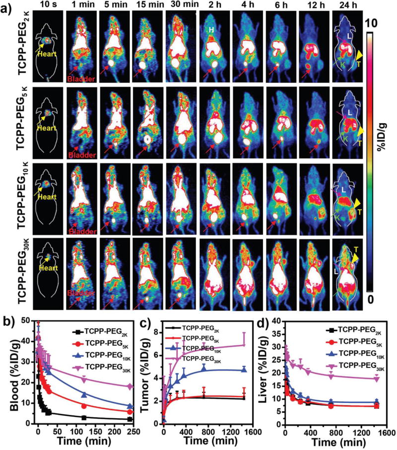
Figure 3.
In vivo PET imaging: a) PET images of 4T1 tumor-bearing mice taken at various time points (10 s, 1 min, 5 min, 15 min, 30 min, 2 h, 4 h, 6 h, 12 h, and 24 h) post-injection of 64Cu-TCPP-PEG2K, 64Cu-TCPP-PEG5K, 64Cu-TCPP-PEG10K, and 64Cu-TCPP-PEG30K nanoparticles. Liver (L), Kidneys (K), Heart, and Bladder are indicated in each figure. Tumor (T) is indicated by yellow arrowheads. (b–d) Time-activity curves of 64Cu-TCPP-PEG (2K, 5K, 10K, and 30K) in different major organs of (b) blood, (c) tumor, and (d) liver.

