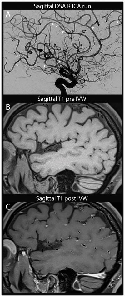
Figure 2.

Reversible cerebral vasoconstriction syndrome with associated enhancement. Sagittal DSA run of the right ICA (A) shows multi-focal stenosis throughout the right MCA, ACA and PCA territories (arrowheads) which was seen in all vascular territories (not shown). On sagittal T1 pre (B) and post-(C) contrast IVWI, there is diffuse arterial wall enhancement with minimal wall thickening. Follow-up CTA at 2 months (not shown) and serial inpatient TCD showed improvement in stenoses.
