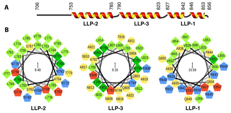
Figure 2. Secondary Structure and Helical Wheel Diagrams of the gp41CTC Protein.
(A) Secondary structure representation of the gp41CT protein based on the NMR data. (B) Helical wheel diagrams of the LLP2, LLP3, and LLP1 motifs. Amino acid sequences are plotted clockwise. Hydrophobic and aromatic residues are represented by light and dark green squares, respectively. Polar residues are shown as yellow circles, while positively and negatively charged residues as blue and red pentagons, respectively. The helical wheels are oriented so that their hydrophobic moments (indicated in the centers) point upwards. Helical wheels were generated via a modified script obtained from http://rzlab.ucr.edu/scripts/wheel/.

