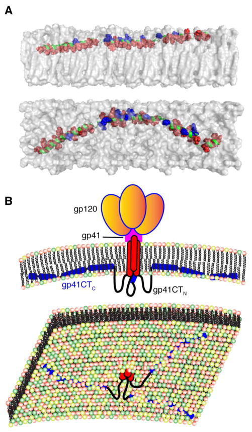
Figure 5. Membrane Interaction of gp41CTC and Overall Env Organization on the Virion Surface.
(A) A model of gp41CTC bound to a membrane bilayer constructed based on the NMR data using the representative structure of gp41CTC with only minor modifications of the dihedral angles in the hinge regions to create an extended molecule. Length of the extended gp41CTC domain shown here is 160 Å. Top and bottom panels show side and top views of the protein, respectively. Residues indicated as red spheres interact extensively with the interior of the membrane while those in blue are mostly exposed and interact with the polar head. Membrane bilayer was generated in VMD membrane builder plug-in (Humphrey et al., 1996). (B) Top panel: A model depicting the gp120 and gp41 proteins on the surface of HIV-1 particles. The gp41CTC domain is penetrating deeply in the inner leaflet of the membrane. Lower panel: An expanded view of the inner leaflet of the PM showing gp41CT penetrating the bilayer.

