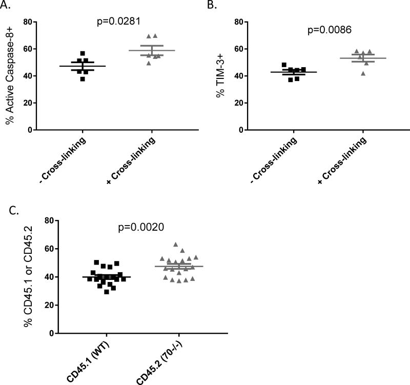
Figure 6. T cell intrinsic CD70 signaling contributes to the inhibitory checkpoint function.
CD4+CD25− T cells were isolated from naïve C57BL/6 WT and CD70−/− mice and added with soluble anti-CD28 to a 48-well plate coated with anti-CD3 the day prior. 0.5×10⁶ T cells were added into each well in 0.5ml medium and treated with IFN-γ at 10ng/ml. 22 hours later the Rat anti-CD70 antibody was added at 5ug/ml to “cross-linking” wells and then 2 hours later a secondary anti-Rat antibody was added to all wells and incubated for another 24 hours. Cells were harvested and analyzed by flow cytometry for active caspase-8 (A) and TIM-3 expression (B). (C) Naïve RAG1−/− mice were injected with 0.5×10⁶ CD4+CD25− T cells purified from CD45.1 mice mixed with 0.5×10⁶ CD4+CD25− T cells from CD45.2 CD70−/− mice. Day 17 after adoptive transfer, spleens were harvested to analyze the percentages of CD45.1 versus CD45.2 cells in the gated TCRβ+CD4+ T cells. Statistical significance was determined by unpaired student t test.

