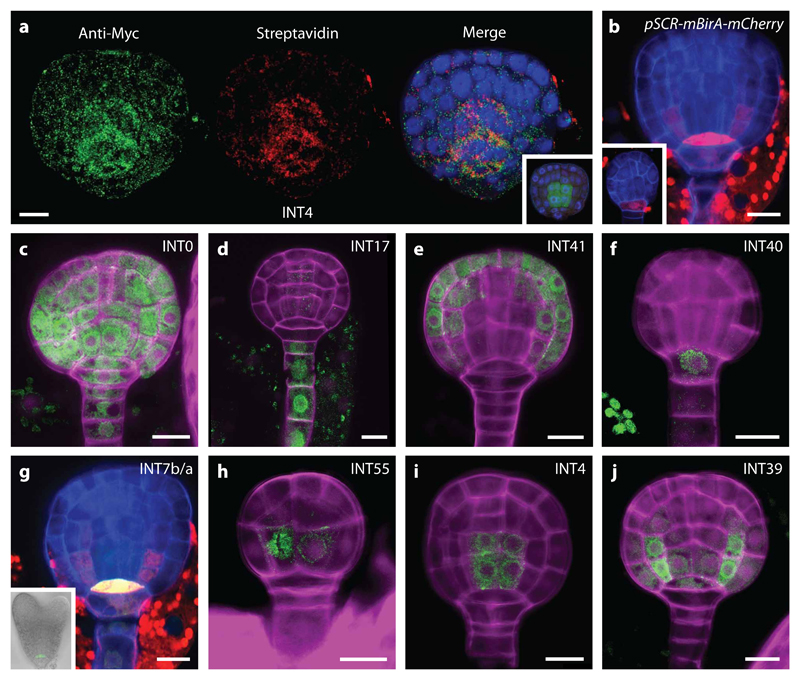
Figure 1. mBirA expression and selected gold-standard INTACT (INT) lines.
(a) Immunostaining of a late globular INT line (INT4; pIQD15-NTF, pWOX2-mBirA-3xMyc) embryo showing whole embryo mBirA expression (Anti-Myc Tag and Alexa Fluor 488 antibody; green) and NTF biotinylation in the vascular tissue precursors (Strepdavidin conjugated with Alexa Fluor 647; red). Counterstaining by DAPI (blue). Insert: negative control (INT4). GFP signal (green) from the pIQD15-NTF is visible in the vascular tissue precursors after fixation. (b) Expression of pSCR-mBirA-mCherry in hypophysis at early globular stage (insert) and in QC and ground tissue precursors at late globular stage. (c-j) INT lines where NTF is expressed in the whole embryo (INT0; c), suspensor (INT17; d), protoderm (INT41; e), hypophysis (INT40; f), QC precursor (INT7b/a; g), and precursor cells (INT55; h) of the vascular (INT4; i) and ground tissue (INT39; j) initials. Scale bar represent 10 μm in all panels.

